The moment after breast cancer surgery marks not an end but a beginning. While the operation itself represents a crucial step in treatment, what happens next to the removed tissue plays an equally vital role in your care journey. Many patients wonder about this mysterious “afterwards” – where does the tissue go? Who looks at it? What information does it provide? The surgical removal of cancerous tissue serves as just one chapter in a longer story that continues in laboratories far from the operating theatre.
The careful examination of removed breast tissue requires expertise and precision. Breast cancer surgeons London rely on detailed pathology reports to make informed decisions about follow-up treatments. When tissue leaves the operating room, it enters a sophisticated process of analysis that provides critical information about your specific cancer. This pathology work often determines whether additional surgery might be needed, what type of medication might work best, and how aggressive your treatment plan should be. The tissue analysis reveals far more than simply confirming cancer – it tells a detailed story about your unique condition.
The methods used for examining breast tissue have evolved significantly over decades, yet rely on fundamental scientific principles that pathologists have refined through years of research and practice. These specialists prepare thin slices of the removed tissue, stain them with special dyes, and examine them under microscopes to identify cancer cells and assess how they differ from normal cells. This meticulous process creates a comprehensive picture of the cancer’s characteristics, helping your medical team understand the specific nature of your condition and determine the most appropriate treatment approach going forward.
The Journey from Operating Theatre to Pathology Lab
The journey begins immediately after surgery. Your surgeon carefully packages the removed tissue, labelling it with your details and relevant surgical information. This specimen travels to the pathology department, typically within the same hospital, where specially trained staff receive and process it. Speed matters here – fresh tissue provides the most accurate results for certain tests, which is why hospitals maintain efficient transport systems between theatres and labs.
Pathology teams first examine the tissue with their naked eyes, a process called gross examination. They note the size, colour, and general appearance of the specimen, taking photographs for reference. They might feel for hard areas that could indicate tumours and measure distances between suspicious areas and surgical margins – the edges of the removed tissue. This seemingly simple step actually provides crucial information about whether the surgery likely removed all cancerous cells.
Slicing and Staining: Preparing Tissue for Analysis
Next comes a fascinatingly precise process. Pathologists cut the specimen into thin sections, sometimes just a few cells thick. These delicate slices get placed on glass slides and stained with dyes that highlight different cellular components. The most common stain, called H&E (haematoxylin and eosin), turns cell nuclei blue-purple and other structures pink, making it easier to identify abnormal cells under the microscope. These stained slides become permanent records that pathologists and oncologists may review multiple times during your treatment.
The detail visible on these slides amazes even experienced medical professionals. Pathologists can see individual cancer cells, their shapes, arrangements, and how they interact with surrounding tissues. They classify the cancer type, grade its aggressiveness, and assess how far it has spread. This microscopic world reveals information impossible to determine during surgery, showing whether cancer cells have invaded blood vessels or lymphatic channels, suggesting potential spread to other body parts.
Beyond Basic Pathology: Specialised Testing
Modern breast cancer care goes further than traditional pathology. Laboratories now conduct sophisticated molecular tests on tissue samples to identify specific genes and proteins that influence how a cancer might behave. Tests like immunohistochemistry determine whether your cancer cells have receptors for hormones (oestrogen and progesterone) or a protein called HER2, which directly affects which treatments might work best for you.
Genetic tests like Oncotype DX or MammaPrint can analyse multiple genes in your cancer cells, predicting how likely the cancer might return and whether chemotherapy would benefit you. These advanced tests sometimes require sending tissue samples to specialised laboratories, explaining why some results take longer to come back than others. The wait feels frustrating, but these detailed analyses provide information that helps avoid unnecessary treatments while ensuring you receive therapies most likely to work.
The Multidisciplinary Team Discussion
After completing all analyses, pathologists compile comprehensive reports detailing their findings. These reports then feature in multidisciplinary team meetings, where surgeons, oncologists, radiologists, pathologists and specialised nurses discuss each patient’s case. The team reviews pathology results alongside your medical history, imaging studies, and other relevant factors to develop personalised treatment recommendations. The tissue analysis forms the foundation for these discussions, providing concrete evidence that guides critical decisions.
This collaborative approach ensures that treatment plans consider all available information. Sometimes, pathology results surprise even experienced clinicians, revealing cancer characteristics that weren’t apparent from scans or clinical examinations. The tissue tells its own story – sometimes reassuring, sometimes challenging – but always providing valuable guidance for the medical team and ultimately for you as the patient.
Understanding Your Pathology Report
When your doctor discusses your pathology report with you, they translate complex medical terminology into understandable information. Key elements typically include the cancer type, its grade (how aggressive it appears), its stage (how far it has spread), hormone receptor status, HER2 status, and whether the surgery achieved “clear margins” meaning no cancer cells were found at the edges of the removed tissue. Each element influences your treatment path moving forward.
Learning to understand your pathology report empowers you to participate more actively in treatment decisions. While the technical language feels overwhelming initially, focusing on how the results affect your specific situation makes the information more manageable. Many breast care nurses excel at explaining these reports, answering questions, and helping you grasp what the findings mean for your particular journey.
What Happens to the Tissue Long-Term?
Most hospitals store tissue samples for many years after surgery, preserving them as part of your medical record. This practice serves several purposes – doctors can re-examine the tissue if questions arise about your diagnosis, researchers might use anonymised samples for cancer studies (with appropriate consent), and new tests developed in the future might provide additional information from these stored samples. This long-term storage explains why you might sign consent forms regarding tissue use during your surgical preparations.
Understanding what happens to breast tissue after surgery helps demystify an important part of cancer care. The detailed analysis performed in pathology laboratories provides crucial information that guides every aspect of your treatment plan. This knowledge empowers you to ask informed questions and better understand the recommendations your medical team makes. If you’ve recently undergone breast cancer surgery or have one scheduled, consider asking your surgeon about the pathology process specific to your hospital – knowing these details might help ease anxiety while waiting for results. Reach out to your breast care nurse with any questions about your pathology report or what it means for your treatment journey ahead.
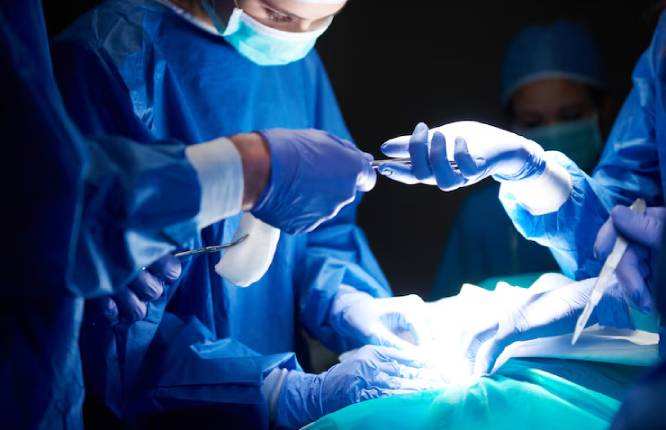
Featured Image Source: https://img.freepik.com/free-photo/surgeons-passing-scissors-each-other_329181-19635.jpg